File:Section through olfactory bulb 16 days old rat brain.jpg
此为最大尺寸。
Section_through_olfactory_bulb_16_days_old_rat_brain.jpg (600 × 356像素,文件大小:205 KB,MIME类型:image/jpeg)
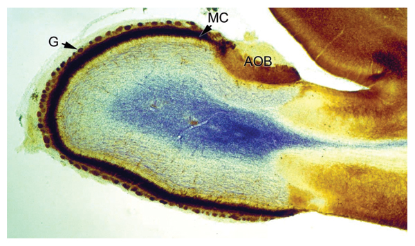
| 描述 | Figure legend from article (CC-by): "Section through the olfactory bulb of a 16 days old rat brain. The tissue has been fixed and immunoperoxidase-stained with antibodies against GABAA-receptor_1-subunit (brown) as described elsewhere [157]. Nissl staining was performed to counter stain (blue). Clearly visible are the accessory olfactory bulb (AOB), to which chemosensory neurons from the vomeronasal organ project, the intensely labelled layer of mitral cells (MC), and the glomeruli (G), which represent the first relay station for sensory information transmitted from the nose to the brain (Jacques Paysan, unpublished)." |
| 日期 | Prepared for Commons on 2007-12-18 |
| 来源 | Elsaesser, Rebecca; Jacques Paysan (2007). "The sense of smell, its signalling pathways, and the dichotomy of cilia and microvilli in olfactory sensory cells". BMC Neuroscience 8 (Suppl 3): S1. DOI:10.1186/1471-2202-8-S3-S1. ISSN 1471-2202. Retrieved on 2007-12-18. |
| 作者 | Prepared for Commons by User:OldakQuill from a CC-by-2.0 figure in a journal article by Rebecca Elsaesser and Jacques Paysan (see source). |
| 授权 (二次使用本文件) |
文件历史
点击某个日期/时间查看对应时刻的文件。
| 日期/时间 | 缩略图 | 大小 | 用户 | 备注 | |
|---|---|---|---|---|---|
| 当前 | 2007年12月18日 (二) 22:51 | | 600 × 356(205 KB) | OldakQuill | {{Information |Description=Figure legend from article (CC-by): "Section through the olfactory bulb of a 16 days old rat brain. The tissue has been fixed and immunoperoxidase-stained with antibodies against GABAA-receptor_1-subunit (brown) as described els |
文件用途
以下页面使用本文件:
全域文件用途
以下其他wiki使用此文件:
- as.wikipedia.org上的用途
- bs.wikipedia.org上的用途
- ceb.wikipedia.org上的用途
- cy.wikipedia.org上的用途
- en.wikipedia.org上的用途
- eo.wikipedia.org上的用途
- es.wikipedia.org上的用途
- fr.wikibooks.org上的用途
- ha.wikipedia.org上的用途
- hy.wikipedia.org上的用途
- outreach.wikimedia.org上的用途
- pl.wikipedia.org上的用途
- pl.wikibooks.org上的用途
- sl.wikipedia.org上的用途
- tr.wikipedia.org上的用途
元数据
🔥 Top keywords: Baike: 首页Special:搜索国际劳动节淚之女王劳动节九龍城寨之圍城2024年湯姆斯盃2024年優霸盃不夠善良的我們背着善宰跑金智媛逆天奇案2春色寄情人金秀賢 (男演員)邊佑錫福建號航空母艦城市猎人 (2024年电影)梅龙高速公路习近平九龍寨城陳耀祥破墓城市猎人笑看風雲六四事件排球少年!!排球少年!!角色列表與鳳行承欢记Seventeen (組合)支配物种劉俊謙 (香港)許瑋甯ILLIT宁安如梦鈴木亮平BABYMONSTER孫綻媽祖中华人民共和国朴成焄周雨彤无用的谎言中華民國張文傑金惠奫周處除三害 (電影)赵长鹏怪獸8號BOYNEXTDOOR李主儐第二十条白鹿 (演員)國道三號崩塌事故澄碧邨乘風2024幕府將軍 (2024年電視劇)哈里·R·杜鲁门李美淑阿努纳奇比利小子特技玩家活塞男事件李现葬送的芙莉蓮IVE (組合)林依晨日本五月天帝國浩劫:美國內戰(G)I-DLEP站末日愚者夜限照相馆三流之路打天下2机动战士GUNDAM SEED FREEDOM張惠東草榴社区三体 (小说)香港鄧麗君迷宮飯NewJeansEnergy (組合)徐巧芯逆天奇案為美好的世界獻上祝福!姜濤搜查班長1958吉伊卡哇張書偉謝京穎艾爾頓·冼拿賀軍翔毛泽东少年歌行轉生為第七王子,隨心所欲的魔法學習之路木村文乃